TELEVECTOR®Lentiviral Vectors
As the fourth generation lentiviral vectors, TELEVECTOR® is developed on the base of the third generation systems. It may effectively integrate the exogenous gene into the host chromosome to achieve a durable and stable expression, at the same time expressing multiple genes and shRNA fragments. The third-generation lentiviral system includes a variety of features that are designed to enhance its biosafety. The correlations with wild-type HIV-1 virus are reduced to a minimum.

Technical Advantages of the TELEVECTOR®
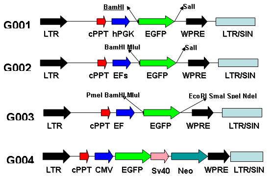
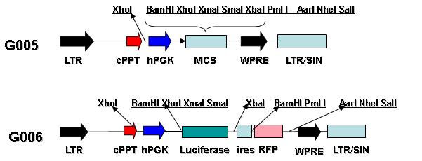
(1) Tailoring to the purpose lentiviral vectors: The optional carrier components include broad-spectrum promoter, tissue-specific expression promoter, red / green / blue fluorescent marker gene, bicistronic, tagged protein and resistance gene and etc..
(2)Wide host range The split and non-split lentiviral vectors from human cells HIV-1 can be active simultaneously infected. The various mammals can be transmitted such as human, rat, mouse, dog, rabbit and etc.. The purpose gene can be delivered to mammalian cells in vitro and in vivo effectively.
(3) The system contains a variety of characteristics that enhance system biosafety. The target gene expression is stable long-term.
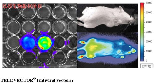
(4) You can choose a reporter gene from GFP, Luciferase, RFP, BFP
The introduction of the reporter gene
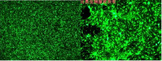
1) EGFP: Enhanced Green Fluorescent Protein is found in jellyfish that is rich in the American northwest coast. It was capable of emitting light for the 65-67 amino acids (Ser - Tyr - Gly) residue in the sequence of 238 amino acids can spontaneously form fluorescent chromospheres.
Luminescence mechanism: when the protein chain folds, amino acid fragment buried deep in the protein will contact closely, leading cyclization and forming imidazolone as well dehydration reaction. If there is molecular oxygen at this time, it can come up with oxidative dehydrogenation. Then it can make the GFP chromophore form emitting fluorescence. GFP is non-toxic, and it can be light-emitting by itself without other coenzyme. When the GFP gene and protein gene which we are interested in fuse, the protein still maintains original activity and does not affect the light-emitting ability. On the fluorescence microscopy, protein location, movement, activity and interaction can observed clearly. Currently, there are a variety of different GFP variants, which further improve the wide application of GFP as genetic markers in biological research.
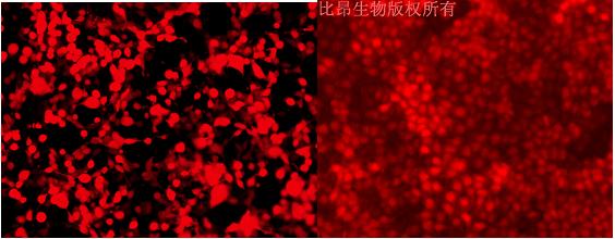
2) RFP:RFP is a dark red fluorescent protein bred by Chudakov greatly improving the quality of intravital imaging and being used widely on the real-time tracking of the molecular activity in the deep tissue of living organisms. Compared with the general fluorescent protein, it can be released out of a longer wavelength so that it can be better used for depth imaging of the live animal offal. And it is helpful for the real-time study of cancer development and treatment process in the form of non-invasive in living organisms, so that we have more in-depth understanding of the pathogenesis of cancer and other diseases. As general fluorescent protein owns weak penetrating, tumors have to be transplanted into subcutaneous shallow or other simulation environments (such as vivisection images or biopsies imaging) to study. Deep red fluorescent proteins may eventually be used in clinical treatment.
3) BFP (blue fluorescent protein): It can be expressed in a variety of cells and products fluorescence spontaneously without other cofactors, which makes it suitable for intravital imaging study. Recently, it has reported that brightness and light sensitivity of Aequorea which is an improved blue fluorescent protein variant as compared with the EBFP are significantly enhanced. These new variants are named as of Azurite, SBFP2 (strongly enhanced blue fluorescent protein and EBFP2 which make it be possible that the living cells in the blue spectral regional imaging for a long time is successful. Even three fluorescent protein show weak dimer characteristics in micro-environment with high concentration, but they can play a great role in the target protein fusion with subcellular localization, as well as they can be imaging easily with standard BFP and 4' ,6-diamidino-2-phenylindole, DAPI through the filter piece of equipment. All these BFP variants can be transformed into a true monomer through A206K mutation that does not affect their characteristics. More importantly, as a blue fluorescent protein EBFP2 with the strongest brightness and the strongest light stability is a good donor of FRET in living cells of EGFP.
4) Luciferase: Bioluminescent organisms are widely distributed in nature: bacteria, fungi, fish, and insects. The enzymes that can catalyze bioluminescence reaction in these bioluminescent organisms are called luciferase, and the substrate is named luciferin.
There is a big difference between luciferase and substrate from different species, but there is one thing in common that there is always the oxidation reaction in the bioluminescent reaction system. Through two steps, there will be the oxidation in the luciferin and luminescent reaction which is ATP-dependent. After adenylation, the heterocycle luciferin products AMP, CO2 by an oxidative decarboxylation and emits light through the activation luciferase intermediate. Mixing the reagents required for the reaction with cell lysate containing the luciferase, the rapid decay (within one second) yellow-green flash (emission peak 560 nm) will appeared. The optical signal can be detected by luminometer equipped with automatic injecting apparatus to facilitate the mixing reactants and can be also recorded by a standard liquid scintillation. When the substrate is excessive, the total amount of light emitting is proportional to the sample fluorescence enzyme activity. Therefore, the transcription of luciferase reporter gene can be estimated indirectly. Luciferase is susceptible to degradation by proteases, and the half-life is approximately 3 hours in the transfection mammalian cells. Luciferase reporter system provides the detection of the promoter activity with sensitive, rapid, non-radioactive detection methods.